Orthopedic implants

Overview

An orthopedic implant is an artificial device targeted to replace a joint, bone, or cartilage damaged or deformed by traumas or diseases. In this case, the doctor may replace a damaged natural part with a manufactured medical device, e.g., an orthopedic implant. However, this task is not easy and heavily depends on the damaged area and types of bones.

Process flow

Computer Tomography (CT), or Magnetic Resonance Imaging (MRI) scannig to gather data on the damaged or deformed bone.

Based on CT data, the damaged bone and the part where the implant should be placed are being modeled.

Plastic implant and the bone are printed. The physical samples is provided to doctor. After feedback and correction the implant is being printed.

The doctor does pre-operation training and optimization of the surgery process. Then, surgery is performed.

Cases

Talus replacement

Case 1: Orbital flaw: Talus

talus_med3-110_51817152527_o-removebg-preview

The story

A young adult with a traumatic fracture-dislocation of the Talus required multiple operations over 2 years. The whole talus developed Avascular Necrosis. Attempted fusion with a locked IM-rod from the calcaneus up the fibia failed. Due to intractable pain, the patient was advised a Syme amputation that he refused. After consulting with our group, understanding the difficulty and possible risk of an ultimate BK amputation, he agreed to get a 3-D printed talar implant specifically designed for him, aiming to achieve a pan-talar fusion including a triple arthodesis with the implant, hoping to keep his foot. The surgery was performed in February 2021. The patient had an uneventful recovery, is now able to walk without pain, full weight bearing on the leg that he retains.

Process of the work

Computer tomography of the damaged bone

3D model of the bone

Designing and modeling of the implant

Planning the surgery

Designing and preparing necessary tools and screws for the surgery.

Preparation of implants

To mitigate the risk 3 different implants are ready for use.

DSC08035

3D printed Titanium implant: 3 different modification of the implants

mediii-4_51019541093_o-removebg-preview
Aren Esayan votq

Damaged talus

Aren Esayan

Implant assembled with damaged bone

Surgery

The surgery was performed in February 2021. The patient had an uneventful recovery. He is now able to walk without pain, full weight bearing on the leg that he retains.

Case 2: Orbital flaw:

H. G., 67 years old Female

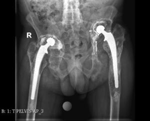
29/10/2015 Right Hip total endoprosthesis performed.

11/05/2016 Left Hip total endoprosthesis performed.

2018 Severe pain in the left thigh and pelvis, swelling of the left thigh. Patient received NSAIDs and Pain Meds to no avail. With ongoing pain, the Left hip was aspirated under CT control, and obtained fluid culture revealed an infection.

02/04/2019 The Left loose and infected femoral stem was revised, lavage, drainage, & a new stem coated with antibiotic cement was inserted. Cup and liner were left in place.

 27/11/2019 Second Revision, removal of all components, insertion of antibiotic spacer.

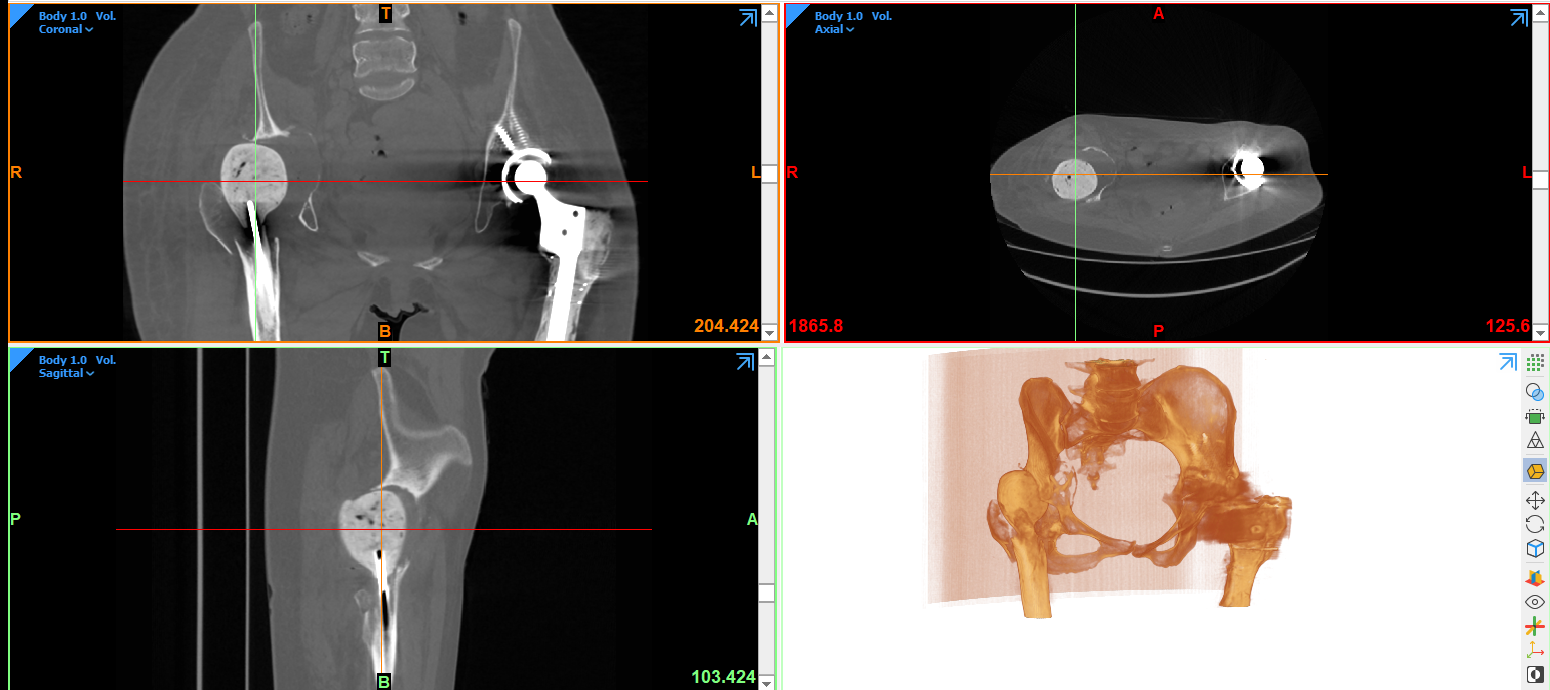
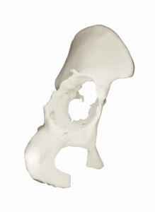
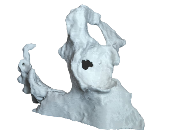
10/09/2020 Removal of the spacer only. Shortening and limp resulted with significant functional as well as acetabular bone deficiency, precluding a standard available component insertion.

[AIP was consulted at this time]

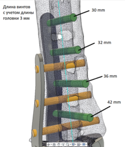
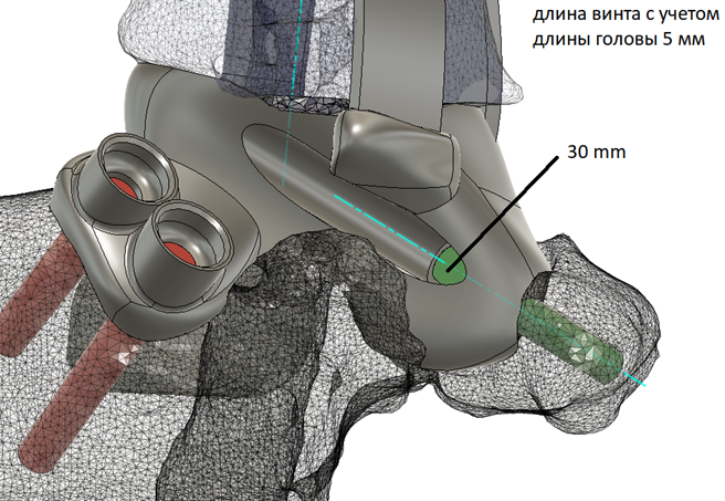
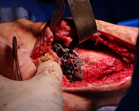
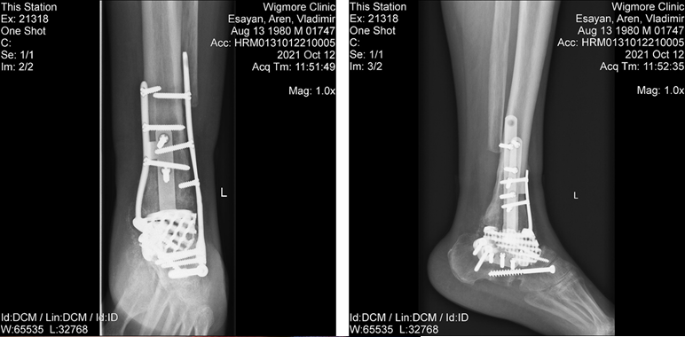
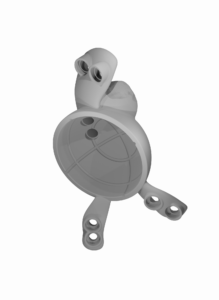
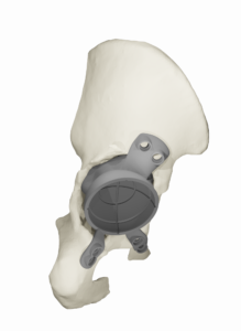
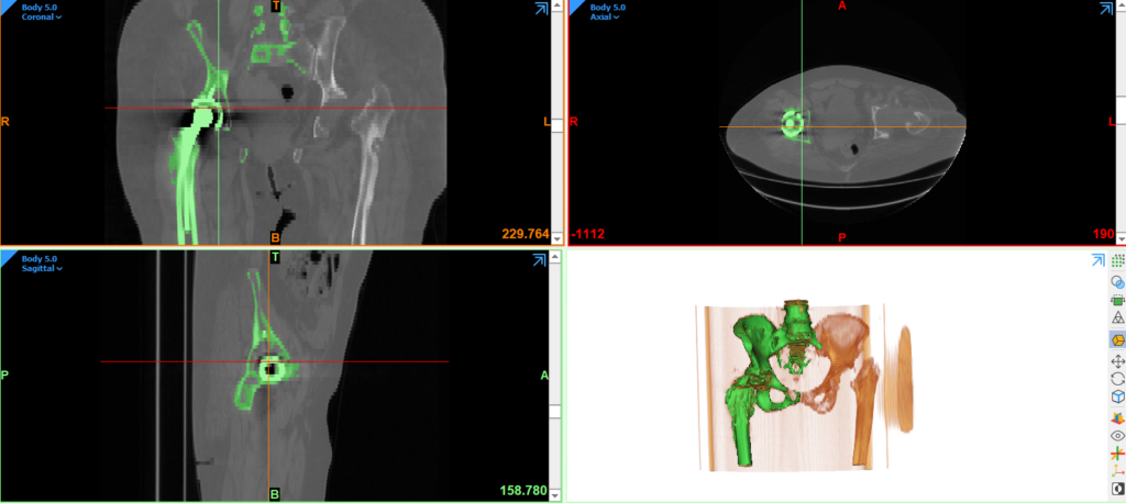
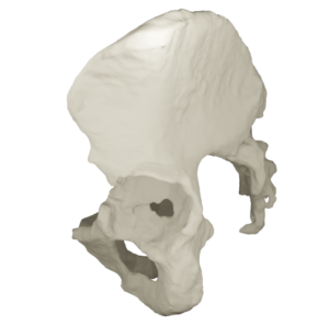
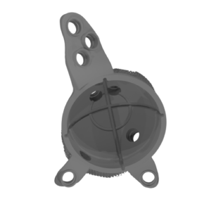
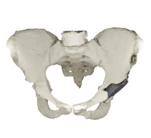
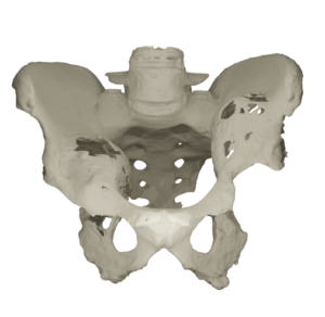
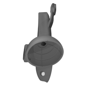
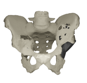
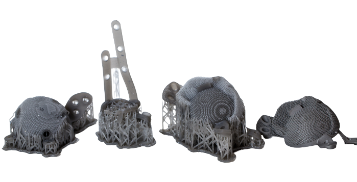
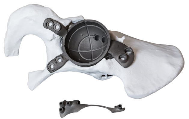
10/03/2021 Final revision was performed with a CT scan based, designed and printed/manufactured acetabular component with a new femoral stem (provided by AIP).

Presently, patient is well, ambulating with equal leg lengths and happy. No recurrence of  pain or infection thus far.

CT and model of the damaged part

Damaged part​

3D printed Titanium implant

Implant assembled with damaged bone

Case 3: Orbital flaw

He is a young beekeeper, in his early 30s. A few years ago he underwent a very difficult and rare surgery, a complete replacement of his two hips. Such a serious operation was necessary since the young man was suffering from a rare disease, which caused the bones to become brittle. Unfortunately, even after 3 surgeries on his left hip and 2 on his right hip, he became disabled, completely incapable of moving and acting on his own, suffering from continuous pain that became a daily companion. He turned to us, seeking salvation, to alleviate the incessant pain, and he was secretly dreaming of perhaps walking again. We immediately embarked on the case․ Such surgery is unique in its complexity in the field of medicine. Large-scale preoperative work began. At first we reconstructed the left hip, designed and modeled from a huge titanium implant to repair the pelvis. We were very satisfied with the result. Next, a custom implant was produced with a 3D printer, after which the patient was successfully operated on. These words do not describe the real complexity and the risks associated with such a difficult operation. The probability of success was very small, but we achieved it. The success was mainly due to the vast and professional preparatory effort that we put into this case. In the second half of the day of the operation, the patient stood up for the first time with some help, however on the next day he started walking on his own.

X-ray image of the patient

Damaged part​

3D printed Titanium implant

Implant assembled with damaged bone

Case 4: Orbital flaw:

The patient was a 30 years old female with a failed total hip replacement. She previously had two failed revisions for infection on this hip. After the infection cleared, the bone defect was so significant and irregular that no standard implant option would have helped her. She lived in pain for several years. Our medical engineers prepared a customized cup for her within two weeks. We are now about six months post-op. So far, the patient is recovering nicely without complications.

CT and model of the damaged part

Thermine Karapetyan2

Damaged part​

3D printed Titanium implant

Implant assembled with damaged bone

Gallery